Photonics for Medical Technologies
Topic Leader: Francesca Rossi
Photonics is one out of 6 broad Key Enabling Technologies (KETs) prioritizing research and innovation in Horizon Europe. Its importance in the Life Science is mainly related to the large availability of light sources, characterized by different wavelengths, emission modalities, beam dimensions: this leads to the design of a tailored solution, thus targeting a specific cell/molecule/tissue and answering a specific medical need with a minimally-invasive device for diagnostics, therapy or treatment.
This concept is at the basis of the research activities performed in the BioPhotonics and Medicine Lab, where the medical need is addressed thanks to the collaboration of experienced international medical staff and companies developing the medical devices.
Among the several research area covered by the group activity within the years, here we focus on laser assisted repair of biological tissues, LEDs application in wound healing, photoaoustics microscopy and sensing, hybrid organosilicon / polyol phantoms for applications in biophotonics
Laser-assisted repair of biological tissues
We proposed new methods of laser-assisted repair of biological tissues (patented) to replace conventional suturing, based on laser induced activation of the endogenous collagen, which behaves like a thermally activated glue. Our method received the approval of the Italian Health Ministry to perform both pre-clinical and clinical experimentations in Ophthalmology and pre-clinical experimentations in brain microsurgery.
A recent optimization consisted in the introduction of new light-responsive hybrid bioadhesives for tissue repair that can be precisely bonded to a biological tissue by photothermal activation. These are easily-handy and resorbable medical dressings that are embedded with laser-activatable organic chromophores or nanochromophores. Laser illumination of the chromophores triggers a strong adhesion between the bioadhesive and the tissue.
For more info please contact Francesca Rossi
Main references:
- Rossi F, Magni G, Colasanti R, Banchelli M, Iacoangeli M, Carrassi E, Aiudi D, Di Rienzo A, Giannoni L, Pieri L, Dallari S, Pini R, Matteini P. (2021). Characterization and Ex Vivo Application of Indocyanine Green Chitosan Patches in Dura Mater Laser Bonding. Polymers 13(13) 2130. https://doi.org/10.3390/polym13132130.
- Alessio Milanesi, Giada Magni Sonia Centi Gioacchino Schifino Annalisa Aluigi Boris N. Khlebtsov Lucia Cavigli Andrea Barucci Nikolai G. Khlebtsov Fulvio Ratto Francesca Rossi Roberto Pini (2020). Optically activated and interrogated plasmonic hydrogels for applications in wound healing. Journal of Biophotonics 13(9) e202000135 https://doi.org/10.1002/jbio.202000135.
- Rossi F, Canovetti A, Malandrini A, Lenzetti I, Pini R, Menabuoni L. (2015). An “All-laser” Endothelial Transplant. J. Vis. Exp. (101), e52939, https://dx.doi.org/10.3791/52939.
- Canovetti A, Malandrini A, Lenzetti I, Rossi F, Pini R, Menabuoni L. (2014) Laser-assisted penetrating keratoplasty: one year’s results in patients, using a laser-welded “anvil”-profiled graft. Am J Ophthalmol. 158(4):664-670.e2. https://doi.org/10.1016/j.ajo.2014.07.010.
- Esposito G, Rossi F, Matteini P, Scerrati A, Puca A, Albanese A, Rossi G, Ratto F, Maira G, Pini R. (2013) In vivo laser assisted microvascular repair and end-to-end anastomosis by means of indocyanine green-infused chitosan patches: A pilot study. Lasers Surg Med., 45(5):318-25. https://doi.org/10.1002/lsm.22145.
- Matteini P, Ratto F, Rossi F, de Angelis M, Cavigli L, Pini R. (2012) Hybrid nanocomposite films for laser-activated tissue bonding. J Biophotonics. 5 (11-12): 868-77. https://doi.org/10.1002/jbio.201200115.
- F. Rossi, P. Matteini, F. Ratto, L. Menabuoni, I. Lenzetti, R. Pini (2008). Laser tissue welding in ophthalmic surgery. J Biophotonics, 1(4), 331-342, https://doi.org/10.1002/jbio.200810028.
LED technologies for photocoagulation and wound healing
We developed new technologies based on the use of high power blue-LEDs to repair abrasions and wounds. The selected wavelength is in the blue region and perfectly matches the absorption peak of the Heme Group and Cytochrome C. The main effects are: a photothermal effect confined in the blood content, providing an immediate coagulation due to the localized temperature rise; a photochemical effect modulating the Cytochrome C. The result is that the device can be used for haemostasis of superficial bleeding wounds and to modulate the activity of cells that are involved in the wound healing process.
For more info please contact Francesca Rossi
Main references:
- Rossi F, Magni G, Tatini F, Banchelli M, Cherchi F, Rossi M, Coppi E, Pugliese AM, Rossi degl’Innocenti D, Alfieri D, Pavone FS, Pini R, Matteini P. (2021). Photobiomodulation of Human Fibroblasts and Keratinocytes with Blue Light: Implications in Wound Healing. Biomedicines. 9(1):41. https://doi.org/10.3390/biomedicines9010041.
- Magni G, Banchelli M, Cherchi F, Coppi E, Fraccalvieri M, Rossi M, Tatini F, Pugliese AM, Rossi Degl’Innocenti D, Alfieri D, Matteini P, Pini R, Pavone FS, Rossi F. (2020) Experimental Study on Blue Light Interaction with Human Keloid-Derived Fibroblasts. Experimental Study on Blue Light Interaction with Human Keloid-Derived Fibroblasts. Biomedicines. 8(12):573. https://doi.org/10.3390/biomedicines8120573.
- Giada Magni, Francesca Tatini, Stefano Bacci, Gaia Paroli, Gaetano De Siena, Riccardo Cicchi, Francesco S. Pavone, Roberto Pini Francesca Rossi (2019) Blue LED light modulates inflammatory infiltrate and improves the healing of superficial wounds. Photodermatology, Photoimmunology &Photomedicine 36(2): 166-168 https://doi.org/10.1111/phpp.12527.
- Cicchi R, Rossi F, Alfieri D, Bacci S, Tatini F, De Siena G, Paroli G, Pini R, Pavone FS. (2016) Observation of an improved healing process in superficial skin wounds after irradiation with a blue-LED haemostatic device. J Biophotonics. 9(6) 645-655 https://doi.org/10.1002/jbio.201500191.
Photoacoustic microscopy and sensing
Photoacoustic microcopy and tomography are emerging as powerful solutions in biomedical diagnostics.
These methods rely on the analysis of ultrasounds that are generated by the rapid photothermal heating and thermoelastic expansion occurring upon absorption of pulsed light. The combination of optical excitation and acoustic detection allows one to reach far deeper penetration into biological tissue than wholly optical imaging techniques, while maintaining the high contrast and spectroscopic specificity of optical imaging. Examples of applications include the interrogation of endogenous dyes as hemoglobin or melanin to map the vascular network or the distribution of melanosomes in various contexts, such as oncology or ophthalmology.
However, the potential of these technologies may readily extend to the molecular imaging sector by the use of molecularly-targeted optical contrast agents.
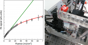
In this context, we developed a custom photoacoustic microscope set-up dedicated to the optimization and characterization of exogenous contrast agents for photoacoustic applications, with a specific focus on plasmonic nanoparticles and gold nanorods. A review of our main results has been reported here
Recently, in the framework of all-optical photoacoustic imaging and sensing, we developed a setup based on whispering-gallery-mode microbubble resonators as a microfluidic platform for material inspection via their photoacoustic response, achieving all-optical detection and high sensitivity towards the material spectral absorbance.
In this setup, the microbubble resonator acts both as the acoustic transducer and as the vial for the sample causing the photoacoustic event, thus dictating its operational frequency and removing any need for impedance-matching media.
For more info please contact Lucia Cavigli
Main references:
- Lucia Cavigli, Boris N. Khlebtsov, Sonia Centi, Nikolai G. Khlebtsov, Roberto Pini and Fulvio Ratto (2021). Photostability of Contrast Agents for Photoacoustics: The Case of Gold Nanorods Nanomaterials 11(1), 116; https://doi.org/10.3390/nano11010116
- Frigenti, G.; Cavigli, L.; Fernández-Bienes, A.; Ratto, F.; Centi, S.; García-Fernández, T.; Nunzi Conti, G.; Soria, S. (2020) Microbubble Resonators for All-Optical Photoacoustics of Flowing Contrast Agents. Sensors 20, 1696. https://doi.org/10.3390/s20061696
- Gabriele Frigenti, Lucia Cavigli, Alberto Fernández-Bienes, Fulvio Ratto, Sonia Centi, Tupak García-Fernández, Gualtiero Nunzi Conti, and Silvia Soria (2019). Resonant Microbubble as a Microfluidic Stage for All-Optical Photoacoustic Sensing. Phys. Rev. Applied 12, 014062 https://doi.org/10.1103/PhysRevApplied.12.014062
- Cavigli, L., Tatini, F., Borri, C., Ratto, F., Centi, S., Cini, A., Lelli, B., Matteini, P., Pini, R. (2016) Preparation and Photoacoustic Analysis of Cellular Vehicles Containing Gold Nanorods. J. Vis. Exp. (111), e53328, https://dx.doi.org/10.3791/53328.
Hybrid organosilicon / polyol phantoms for applications in biophotonics
A growing awareness of the ethical issues posed by in vivo testing is pushing for new technologies for replacing, reducing and refining the use of lab animals for translational research. In this context, the use of anatomical phantoms is a well-established alternative of particular relevance in many situations where the objective assessment of system performances is the principal need. Meanwhile, recent advances of medical technologies toward hybrid and multi-modality are demanding a new generation of phantom materials that may encode complex combinations of the biophysical features of bio-tissue. Examples include hybrid methods like photoacoustic imaging, which involves an interplay of optical, acoustic and thermodynamic features, or multimodal protocols like MRI-guided photothermal and radiation treatments.
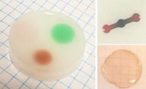
In this framework, we are developing composite materials that may account for the ultrastructural and biochemical complexity of bio-tissue, i.e. accommodate a broad variety of endogenous and exogenous agents of biophysical contrast and enable high fidelity of anatomical reconstruction over multiple length scales. Our long-term vision is to produce a resilient and ethical technology that may replicate the interactions between bio-tissue and physical agents of any kind.
For more info please contact Fulvio Ratto or Lucia Cavigli
Main references:
- Lucia Cavigli, Sonia Centi, Claudia Borri, Giada Magni, Andrea Barucci, Marina Mazzoni, Roberto Carpi, Roberto Incalcaterra, Giacomo Belli, Roberto Pini, Fulvio Ratto, (2020) Hybrid organosilicon/polyol phantoms for applications in biophotonics and beyond, Proc. SPIE 11231, Design and Quality for Biomedical Technologies XIII, 112310S https://doi.org/10.1117/12.2545002
- Fulvio Ratto, Lucia Cavigli, Claudia Borri, Sonia Centi, Giada Magni, Marina Mazzoni, and Roberto Pini, (2019) Hybrid organosilicon/polyol phantom for photoacoustic imaging, Biomed. Opt. Express 10, 3719-3730 https://doi.org/10.1364/BOE.10.003719
- Cinzia Avigo, Nicole Di Lascio, Paolo Armanetti, Claudia Kusmic, Lucia Cavigli, Fulvio Ratto, Sandro Meucci, Cecilia Masciullo, Marco Cecchini, Roberto Pini, Francesco Faita, Luca Menichetti, (2015) Organosilicon phantom for photoacoustic imaging, J. Biomed. Opt. 20(4) 046008 https://doi.org/10.1117/1.JBO.20.4.046008
Current members:
- Francesca Rossi (topic leader)
- Andrea Barucci
- Claudia Borri
- Lucia Cavigli
- Sonia Centi
- Giada Magni
- Paolo Matteini
- Filippo Micheletti
- Roberto Pini
- Fulvio Ratto